Figure 6 from Femoral Hernia: A Review of the Clinical Anatomy and
$ 12.00 · 4.9 (74) · In stock

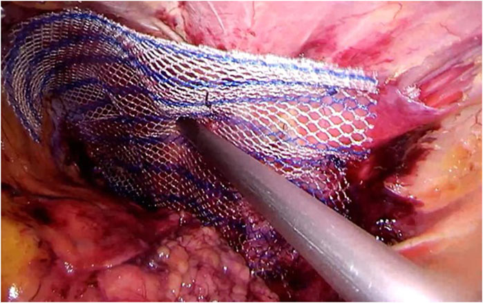
Figure 6. Femoral hernia repair in clean operation. (a) The narrow side of the mesh is sutured to Cooper’s ligament; (b) The mesh is sutured to the iliopubic tract or shelving portion of the inguinal ligament; (c) The posterior wall of the inguinal canal is reinforced, as in Lichtenstein’s repair. - "Femoral Hernia: A Review of the Clinical Anatomy and Surgical Treatment"
Frontiers Publishing Partnerships Primary Lumbar Hernia, Review and Proposals for a Standardized Treatment
Embryonic developmental process and clinical anatomy of the preperitoneal fascia and its clinical significance
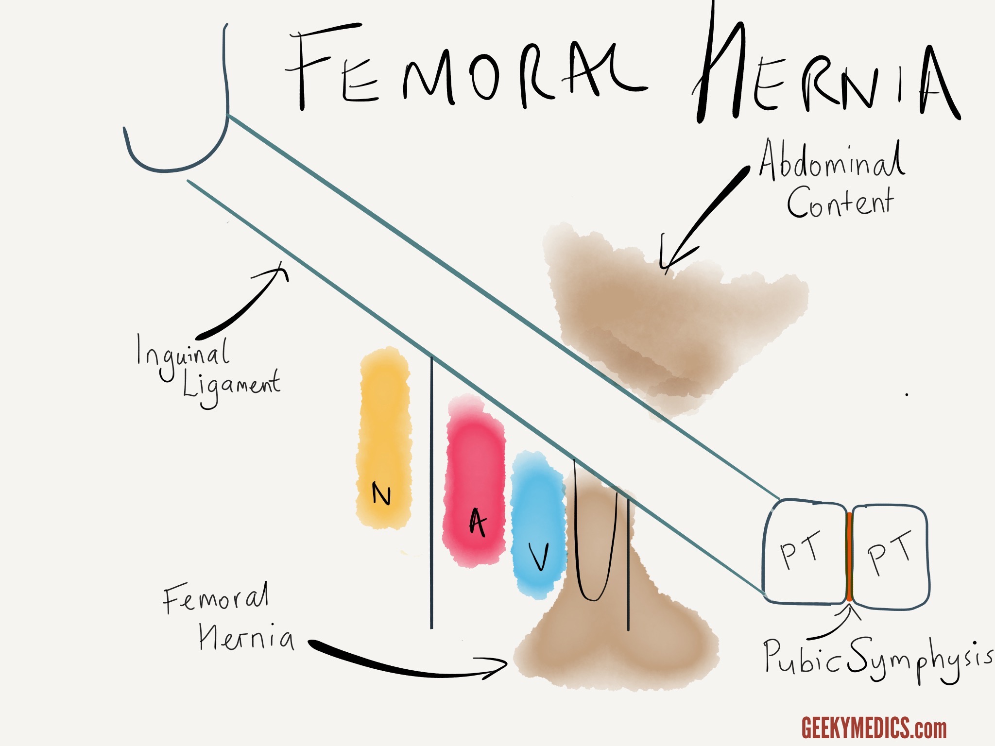
Femoral Region and Hernias: Anatomy - Lecturio Medical
Cureus, Femoral Hernia Containing a Strangulated Appendix: A Hybrid Approach
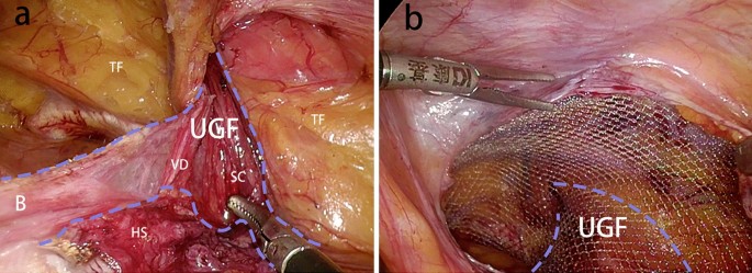
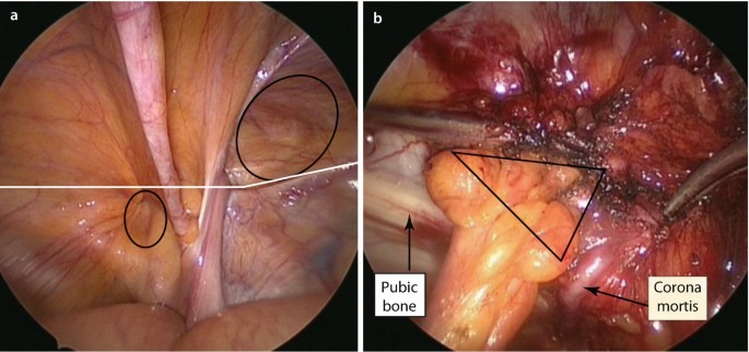
8 Anatomical basis of the myopectineal orifice (Fruchaud) or inner

Figure 7 from Femoral Hernia: A Review of the Clinical Anatomy and Surgical Treatment
Myopectineal orifice. The oval-shaped myopectineal orifice (green
Hernias, Inguinal, Femoral, Umbilical

Figure 2 from Femoral Hernia: A Review of the Clinical Anatomy and Surgical Treatment

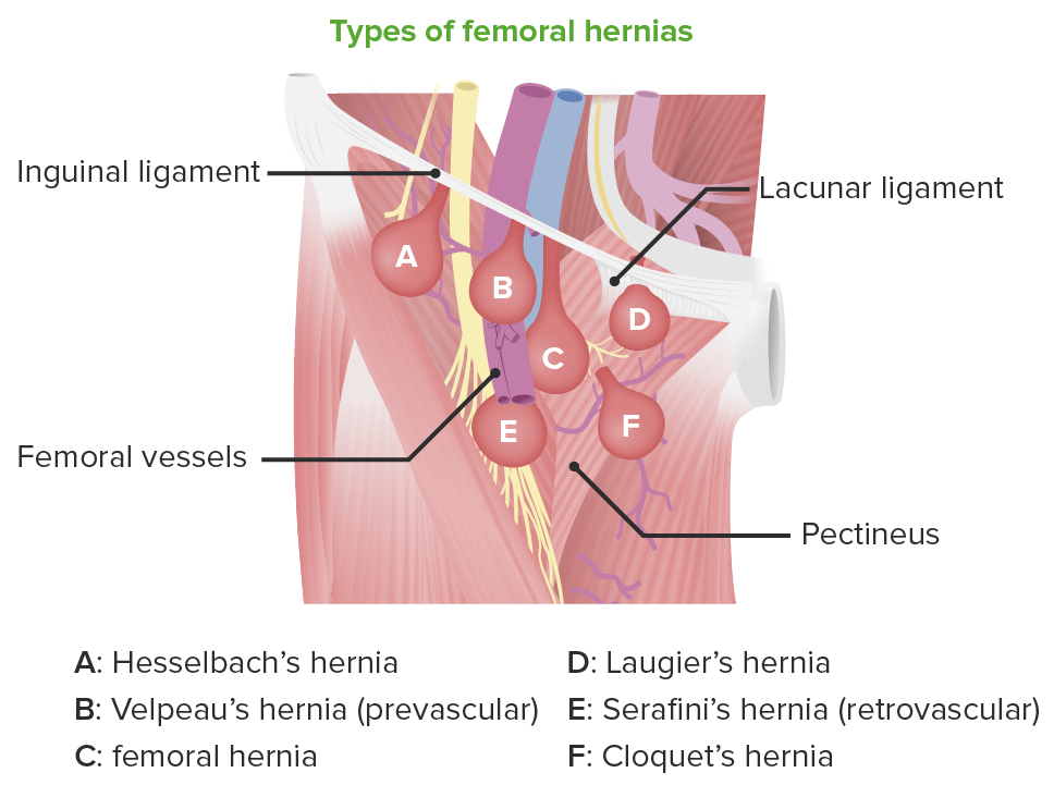
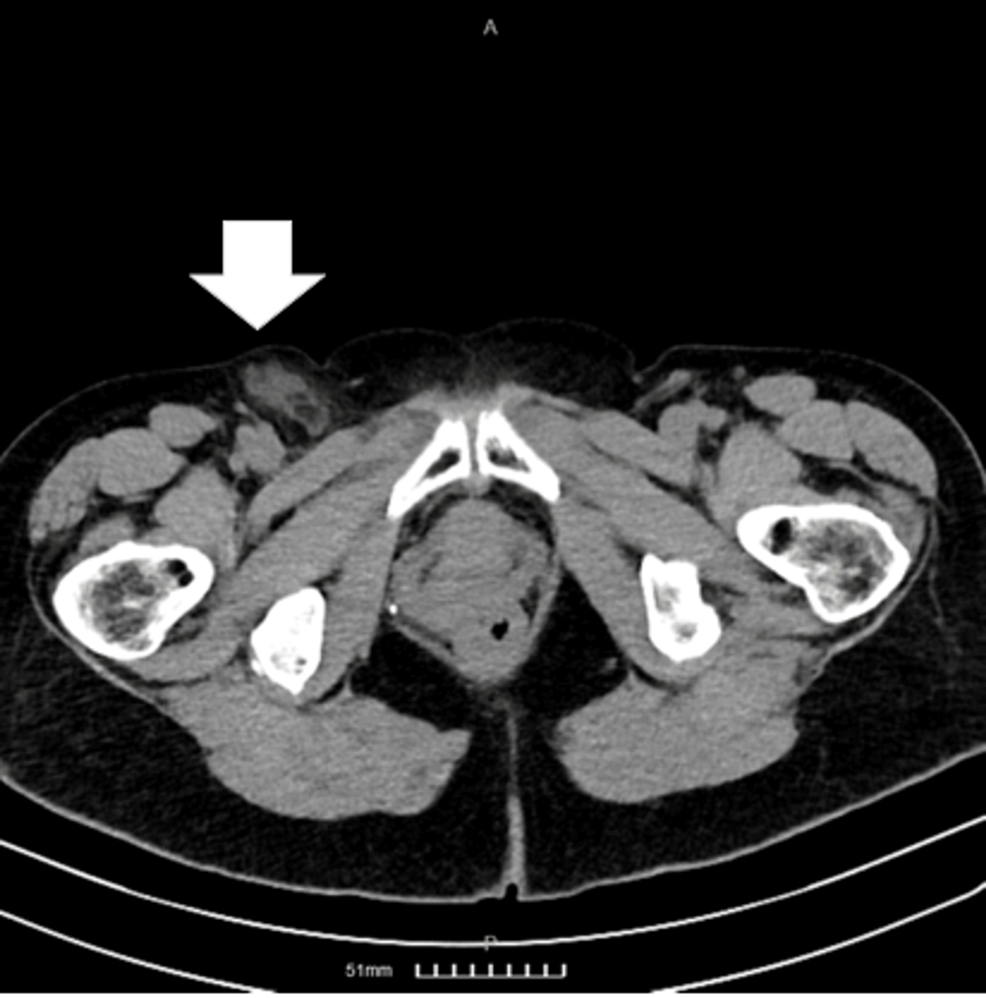
Right incarcerated femoral hernia; the contents of the hernia were the
Clinical Anatomy of the Groin: Posterior Laparoscopic Approach

Anterior and posterior views of myopectineal orifice ( from Elliott and

Femoral Hernia - A Review of Clinical Anatomy
Surgical Techniques Development, Free Full-Text

Diagnostics, Free Full-Text