Pitfalls of inferior vena cava M-mode – NephroPOCUS
$ 7.99 · 4.8 (382) · In stock

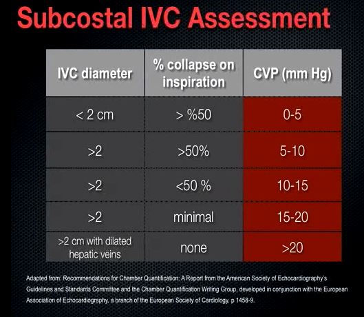
Visual estimation of IVC collapse on B-mode (grey scale image) is generally preferred to M-mode, though in theory, M-mode measurement might be able to give accurate collapsibility index. There are several reasons for this. A major limitation of IVC M-mode is that the vessel moves mediolaterally and craniocaudally during respiration, with collapse occurring off axis…

Point-of-care ultrasound in diagnosis and management of congestive
JCM, Free Full-Text

Venous Excess Doppler Ultrasound for the Nephrologist: Pearls and Pitfalls - ScienceDirect

Image Acquisition Method for the Sonographic Assessment of the

Ultrasonography in Acute Kidney Injury – POCUS Journal

Thread by @NephroP on Thread Reader App – Thread Reader App

Cardiac – Page 2 – NephroPOCUS
Transcending boundaries: Unleashing the potential of multi-organ point-of-care ultrasound in acute kidney injury
The RUSH Ultrasound Exam by Critical Care Specialist Liz Turner

Changes in the Inferior Vena Cava Are More Sensitive Than Venous